Virtaussytometria
Johdanto
Virtaussytometria on menetelmä, jolla voidaan nopeasti havaita ja mitata yksittäisten solujen tai hiukkasten fysikaalisia ja kemiallisia ominaisuuksia, kun ne liuoksessa kulkevat yhden tai useamman laserin valonsäteen läpi. Solut tai hiukkaset havaitaan virtaussytometrin antureilla niiden valon hajontaan ja fluoresenssisäteilyyn perustuvien signaalien perusteella, jotka muunnetaan sähköisiksi signaaleiksi, joita voidaan visualisoida ja analysoida määrällisesti reaaliajassa tietokoneella. Tämä tuottaa tietoa solujen pinnan ja soluliman molekyyleistä sekä solun koosta ja rakenteesta, mikä tekee virtaussytometriasta erinomaisen työkalun solujen ominaisuuksien ja toiminnan analysointiin. Solujen havaitsemisessa apuna käytetään tyypillisesti fluoresenssikonjugoituja vasta-aineita, elävyysvärejä tai DNA:ta sitovia aineita tai solujen ilmentämiä fluoresoivia proteiineja. Yleisesti käytettyjä virtaussytometrian sovelluksia ovat biomarkkereiden analysointi, solusykli, apoptoosi, proteiinien fosforylaatio, kalsiumin virtaus, DNA ja mRNA, spesifinen ligandien sitoutuminen sekä mikro-organismien havaitseminen. Virtaussytometria tunnetaan parhaiten immunologian sovelluksistaan, mutta se on tehokas työkalu myös molekyylibiologiassa, virologiassa, bakteeriologiassa, syöpätutkimuksessa ja sitä käytetään laajalti diagnostiikassa. Solunlajittelusytometri pystyy erottamaan ja puhdistamaan kiinnostavat solupopulaatiot jatkokäyttöä varten. Solujen lajittelu perustuu samoihin ominaisuuksiin, joita lajittelusytometri voi analysoida.
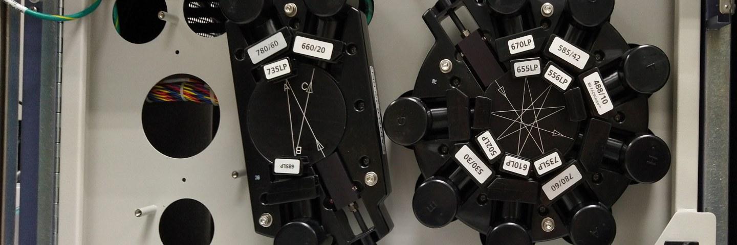
Virtaussytometriapalvelun laitteisto sisältää neljä virtaussytometriä ja yhden aineenvaihdunnan analysoijan, jotka palvelevat eri tutkijoiden tarpeita. Kaikki laitteet soveltuvat analyysiin, ja kaksi niistä ovat myös lajittelijoita. Yksi lajittelija sijaitsee Virus-tilassa. Laitteissa on 1-4 laseria ja ne voivat analysoida jopa 16 erilaista fluoresenssiparametria samanaikaisesti. Käyttäjät voivat käyttää laitteita itse tai vaihtoehtoisesti tutkijat voivat ostaa tarvitsemansa palvelun laitoksen henkilökunnalta. Laitos tarjoaa laajan ja joustavan valikoiman tutkimuspalveluita, aina paneelin suunnittelusta näytekäsittelyapuun ja saadun datan jälkianalyysiin. Yksityiskohtia ja tiedusteluja voi kysyä virtaussytometrian palvelujen johtajalta.
Varausjärjestelmä
Kiitokset
Kaikki käyttäjät ovat velvollisia mainitsemaan virtaussytometriayksikön (Tampere University Flow Cytometry Facility, TFCF) ja Biokeskus Suomen (Biocenter Finland, BF) julkaisuissaan:
“The authors acknowledge Biocenter Finland (BF) and Tampere University Flow Cytometry Facility (TFCF) for their service.”
Laitteet
Virtaussytometrien ja aineenvaihdunnan analysoijan käyttö on sallittu vain kattavan perehdytyksen jälkeen, jota valvovat laitteiden vastuuhenkilöt (ks. Yhteystiedot). Opastus voidaan räätälöidä tutkijoiden yksilöllisten tarpeiden ja aiemman kokemuksen mukaan; kunkin laitteen osalta löytyy esimerkkejä hinnoittelusta. Peruslaboratoriotarvikkeet ja 5 ml 12x75 mm putket, jotka soveltuvat lajitteluun ja analyysiin, sisältyvät käyttömaksuihin. Viratussytometrian palvelu tarjoaa reagenssit ja liuokset, jotka tarvitaan laitteiden asetuksiin ja toimintaan. Näytepuskurit ja värjäysreagenssit on käyttäjien itsensä hankittava. Laitteiden käytön laskutus perustuu Agendo-varauksiin.
Beckman Coulter CytoFlex S
BD FACSCanto II
BD FACSAria Fusion
BD FACSMelody
BD FlowJo Software
Agilent Seahorse XFe24 Analyzer
Valid from 1.11.2025
| CytoFlex S | Price |
| Analysis | 18 €/h |
| Tutorial (2 h)* | 146 € |
| FACSAria Fusion | Price |
| Analysis only | 21 €/h |
| Sorting | 24 €/h |
| Sorting tutorial (3x5 h)* | 1185 € |
| FlowJo software license | Price |
| University group license | 354 €/year |
| Assistance | Price |
| Assistance/service, scheduled beforehand** | 55€/h |
*Tutorials include instrument and service fees. Additional hours will be charged for both.
**Scheduled service or assistance refers to eg. sorting or analysis service or extra training.
Pricing is valid for user groups from University of Tampere. Other academic or non-academic users are asked to inquire specific pricing.
The Flow Cytometry Facility reserves the right to price adjustments.
Julkaisut
2025
Uusi-Mäkelä M, Harjula SE, Junno M, Sillanpää A, Nätkin R, Niskanen MT, Saralahti AK, Nykter M, Rämet M. The inflammasome adaptor pycard is essential for immunity against Mycobacterium marinum infection in adult zebrafish. Dis Model Mech. 2025 Sep 1;18(9):dmm052061. doi: 10.1242/dmm.052061. Epub 2025 Mar 24.
Gaertner K, Tapanainen R, Saari S, Fekete Z, Goffart S, Pohjoismäki JLO, Dufour E. Exploring mitonuclear interactions in the regulation of cell physiology: Insights from interspecies cybrids. Exp Cell Res. 2025 Mar 15;446(2):114466. doi: 10.1016/j.yexcr.2025.114466. Epub 2025 Feb 18.
Hermelo I, Virtanen T, Salonen I, Nätkin R, Keitaanniemi S, Tiihonen AM, Lehtipuro S, Kummola L, Raulamo E, Nordfors K, Haapasalo H, Rauhala M, Kesseli J, Nykter M, Haapasalo J, Rautajoki K. Unsupervised clustering reveals noncanonical myeloid cell subsets in the brain tumor microenvironment. Cancer Immunol Immunother. 2025 Jan 3;74(2):63. doi: 10.1007/s00262-024-03920-1.
2024
Virtanen A, Kettunen V, Musta K, Räkköläinen V, Knapp S, Haikarainen T, Silvennoinen O. Molecular basis of JAK kinase regulation guiding therapeutic approaches: Evaluating the JAK3 pseudokinase domain as a drug target. Adv Biol Regul. 2025 Jan;95:101072. doi: 10.1016/j.jbior.2024.101072. Epub 2024 Dec 24.
Gaertner K, Terzioglu M, Michell C, Tapanainen R, Pohjoismäki J, Dufour E, Saari S. Species differences in glycerol-3-phosphate metabolism reveals trade-offs between metabolic adaptations and cell proliferation.Biochim Biophys Acta Bioenerg. 2024 Dec 2;1866(2):149530. doi: 10.1016/j.bbabio.2024.149530. Epub ahead of print.
Salminen TS, Vesala L, Basikhina Y, Kutzer M, Tuomela T, Lucas R, Monteith K, Prakash A, Tietz T, Vale PF. A naturally occurring mitochondrial genome variant confers broad protection from infection in Drosophila. PLoS Genet. 2024 Nov 11;20(11):e1011476. doi: 10.1371/journal.pgen.1011476.
Lampinen V, Ojanen MJT, Caro FM, Gröhn S, Hankaniemi MM, Pesu M, Hytönen VP. Experimental VLP vaccine displaying a furin antigen elicits production of autoantibodies and is well tolerated in mice. Nanoscale Adv. 2024 Oct 9;6(24):6239–52. doi: 10.1039/d4na00483c. Epub ahead of print.
Yrjänäinen A, Mesiä E, Lampela E, Kreutzer J, Vihinen J, Tornberg K, Vuorenpää H, Miettinen S, Kallio P, Mäki AJ. Barrier-free, open-top microfluidic chip for generating two distinct, interconnected 3D microvascular networks. Sci Rep. 2024 Oct 2;14(1):22916. doi: 10.1038/s41598-024-74493-3.
Miao Y, Virtanen A, Zmajkovic J, Hilpert M, Skoda RC, Silvennoinen O, Haikarainen T. Functional and Structural Characterization of Clinical-Stage Janus Kinase 2 Inhibitors Identifies Determinants for Drug Selectivity. J Med Chem. 2024 Jun 27;67(12):10012-10024. doi: 10.1021/acs.jmedchem.4c00197. Epub 2024 Jun 6.
Vuorenpää H, Valtonen J, Penttinen K, Koskimäki S, Hovinen E, Ahola A, Gering C, Parraga J, Kelloniemi M, Hyttinen J, Kellomäki M, Aalto-Setälä K, Miettinen S, Pekkanen-Mattila M. Gellan gum-gelatin based cardiac models support formation of cellular networks and functional cardiomyocytes. Cytotechnology. 2024 Aug;76(4):483-502. doi: 10.1007/s10616-024-00630-5. Epub 2024 May 2.
Vesala L, Basikhina Y, Tuomela T, Nurminen A, Siukola E, Vale PF, Salminen TS. Mitochondrial perturbation in immune cells enhances cell-mediated innate immunity in Drosophila. BMC Biol. 2024 Mar 13;22(1):60. doi: 10.1186/s12915-024-01858-5.
2023
Zapilko V, Moisio S, Parikka M, Heinäniemi M, Lohi O. Generation of a Zebrafish Knock-In Model Recapitulating Childhood ETV6::RUNX1-Positive B-Cell Precursor Acute Lymphoblastic Leukemia. Cancers (Basel). 2023 Dec 13;15(24):5821. doi: 10.3390/cancers15245821.
Peussa H, Fedele C, Tran H, Marttinen M, Fadjukov J, Mäntylä E, Priimägi A, Nymark S, Ihalainen TO. Light-Induced Nanoscale Deformation in Azobenzene Thin Film Triggers Rapid Intracellular Ca2+ Increase via Mechanosensitive Cation Channels. Adv Sci (Weinh). 2023 Dec;10(35):e2206190. doi: 10.1002/advs.202206190. Epub 2023 Nov 9.
Suominen S, Hyypijev T, Venäläinen M, Yrjänäinen A, Vuorenpää H, Lehti-Polojärvi M, Räsänen M, Seppänen A, Hyttinen J, Miettinen S, Aalto-Setälä K, Viiri LE. Improvements in Maturity and Stability of 3D iPSC-Derived Hepatocyte-like Cell Cultures. Cells. 2023 Sep 27;12(19):2368. doi: 10.3390/cells12192368.
Kummola L, González-Rodríguez MI, Marnila P, Nurminen N, Salomaa T, Hiihtola L, Mäkelä I, Laitinen OH, Hyöty H, Sinkkonen A, Junttila IS. Comparison of the effect of autoclaved and non-autoclaved live soil exposure on the mouse immune system : Effect of soil exposure on immune system. BMC Immunol. 2023 Sep 9;24(1):29. doi: 10.1186/s12865-023-00565-0.
Ilmarinen T, Vattulainen M, Kandhavelu J, Bremond-Gignac D, Aberdam D, Skottman H. Production and limbal lineage commitment of aniridia patient-derived induced pluripotent stem cells. Stem Cells. 2023 Aug 26:sxad067. doi: 10.1093/stmcls/sxad067.
Mäntylä M, Montonen T, Azzari L, Mattola S, Hannula M, Vihinen-Ranta M, Hyttinen J, Vippola M, Foi A, Nymark S, Ihalainen TO. Iterative immunostaining combined with expansion microscopy and image processing reveals nanoscopic network organization of nuclear lamina. Mol Biol Cell. 2023 Aug 1;34(9):br13. doi: 10.1091/mbc.E22-09-0448.
Isosaari L, Vuorenpää H, Yrjänäinen A, Kapucu FE, Kelloniemi M, Pakarinen TK, Miettinen S, Narkilahti S. Simultaneous induction of vasculature and neuronal network formation on a chip reveals a dynamic interrelationship between cell types. Cell Commun Signal. 2023 Jun 14;21(1):132. doi: 10.1186/s12964-023-01159-4.
Danielsson BE, Abraham BG, Mäntylä E, Cabe JI, Mayer CR, Rekonen A, Ek F, Conway DE, Ihalainen TO. Nuclear lamina strain states revealed by intermolecular force biosensor. Nat Commun 14, 3867 (2023). https://doi.org/10.1038/s41467-023-39563-6
Mäki-Opas I, Hämäläinen M, Moilanen LJ, Sood H, Leppänen T, Kummola L, Junttila IS, Lehtimäki L, Moilanen E. TRPA1 Mediates Contact Hypersensitivity Induced by 2,4-Dinitrochlorobenzene. J Invest Dermatol. 2023 Jun;143(6):1104-1108.e4. doi: 10.1016/j.jid.2022.12.014.
Salomaa T, Kummola L, González-Rodríguez MI, Hiihtola L, Järvinen TAH, Junttila IS. Low IL-13Rα1 Expression on Mast Cells Tunes Them Unresponsive to IL-13. J Leukoc Biol. 2023 May 24:qiad065. doi: 10.1093/jleuko/qiad065.
Virtanen A, Palmroth M, Liukkonen S, Kurttila A, Haikarainen T, Isomäki P, Silvennoinen O. In vitro profiling of rheumatic-disease-evaluated JAK inhibitors demonstrate differences in JAK isoform selectivity between different types of inhibitors. Arthritis Rheumatol. 2023 May 3. doi: 10.1002/art.42547.
Mahmoud M, Juntunen M, Adnan A, Kummola L, Junttila IS, Kelloniemi M, Tyrväinen T, Huhtala H, Abd El Fattah AI, Amr K, El Erian AM, Patrikoski M, Miettinen S. Immunomodulatory Functions of Adipose Mesenchymal Stromal/Stem Cell Derived From Donors With Type 2 Diabetes and Obesity on CD4 T Cells. Stem Cells. 2023 May 15;41(5):505-519. doi: 10.1093/stmcls/sxad021.
Kummola L, Salomaa T, Ortutay Z, Savan R, Young HA, Junttila IS. IL-4, IL-13 and IFN-γ -induced genes in highly purified human neutrophils. Cytokine. 2023 Apr;164:156159. doi: 10.1016/j.cyto.2023.156159.
Ojanen MJT, Caro FM, Aittomäki S, Ploquin MJ, Ortutay Z, Pekkarinen M, Kesseli J, Vähätupa M, Määttä J, Nykter M, Junttila IS, Järvinen TAH, O Shea JJ, Biron CA, Pesu M. FURIN regulates cytotoxic T-lymphocyte effector function and memory cell transition in mice. Eur J Immunol. 2023 Apr 4:e2250246. doi: 10.1002/eji.202250246. Epub ahead of print. PMID: 37015057.
2022
Kukkonen K, Autio-Kimura B, Rauhala H, Kesseli J, Nykter M, Latonen L, Visakorpi T. Nonmalignant AR-positive prostate epithelial cells and cancer cells respond differently to androgen. Endocr Relat Cancer. 2022 Nov 7;29(12):717-733. doi: 10.1530/ERC-22-0108. PMID: 36219867; PMCID: PMC9644224.
Gaertner K, Michell C, Tapanainen R, Goffart S, Saari S, Soininmäki M, Dufour E, Pohjoismäki JLO. Molecular phenotyping uncovers differences in basic housekeeping functions among closely related species of hares (Lepus spp., Lagomorpha: Leporidae). Mol Ecol. 2022 Nov 1. doi: 10.1111/mec.16755. Epub ahead of print. PMID: 36320183.
Laukkanen S, Veloso A, Yan C, Oksa L, Alpert EJ, Do D, Hyvärinen N, McCarthy K, Adhikari A, Yang Q, Iyer S, Garcia SP, Pello A, Ruokoranta T, Moisio S, Adhikari S, Yoder JA, Gallagher K, Whelton L, Allen JR, Jin AH, Loontiens S, Heinäniemi M, Kelliher M, Heckman CA, Lohi O, Langenau DM. Therapeutic targeting of LCK tyrosine kinase and mTOR signaling in T-cell acute lymphoblastic leukemia. Blood. 2022 Oct 27;140(17):1891-1906. doi: 10.1182/blood.2021015106.
Raivola J, Dini A, Karvonen H, Piki E, Salokas K, Niininen W, Kaleva L, Zhang K, Arjama M, Gudoityte G, Seashore-Ludlow B, Varjosalo M, Kallioniemi O, Hautaniemi S, Murumägi A, Ungureanu D. Multiomics characterization implicates PTK7 in ovarian cancer EMT and cell plasticity and offers strategies for therapeutic intervention. Cell Death Dis. 2022 Aug 17;13(8):714. doi: 10.1038/s41419-022-05161-5.
Lotila J, Hyvärinen T, Skottman H, Airas L, Narkilahti S, Hagman S. Establishment of a human induced pluripotent stem cell line (TAUi008-A) derived from a multiple sclerosis patient. Stem Cell Res. 2022 Aug;63:102865. doi: 10.1016/j.scr.2022.102865.
Raivola J, Dini A, Salokas K, Karvonen H, Niininen W, Piki E, Varjosalo M, Ungureanu D. New insights into the molecular mechanisms of ROR1, ROR2, and PTK7 signaling from the proteomics and pharmacological modulation of ROR1 interactome. Cell Mol Life Sci. 2022 May 4;79(5):276. doi: 10.1007/s00018-022-04301-6.
Oksa L, Mäkinen A, Nikkilä A, Hyvärinen N, Laukkanen S, Rokka A, Haapaniemi P, Seki M, Takita J, Kauko O, Heinäniemi M, Lohi O. Arginine Methyltransferase PRMT7 Deregulates Expression of RUNX1 Target Genes in T-Cell Acute Lymphoblastic Leukemia. Cancers (Basel). 2022 Apr 26;14(9):2169. doi: 10.3390/cancers14092169.
Mykuliak A, Yrjänäinen A, Mäki AJ, Gebraad A, Lampela E, Kääriäinen M, Pakarinen TK, Kallio P, Miettinen S, Vuorenpää H. Vasculogenic Potency of Bone Marrow- and Adipose Tissue-Derived Mesenchymal Stem/Stromal Cells Results in Differing Vascular Network Phenotypes in a Microfluidic Chip. Front Bioeng Biotechnol. 2022 Feb 8;10:764237. doi: 10.3389/fbioe.2022.764237.
2021
Järvelä-Stölting M, Vesala L, Maasdorp MK, Ciantar J, Rämet M, Valanne S. Proteasome α6 Subunit Negatively Regulates the JAK/STAT Pathway and Blood Cell Activation in Drosophila melanogaster. Front Immunol. 2021 Dec 22;12:729631.
Vattulainen M, Ilmarinen T, Viheriälä T, Jokinen V, Skottman H. Corneal epithelial differentiation of human pluripotent stem cells generates ABCB5+ and ∆Np63α+ cells with limbal cell characteristics and high wound healing capacity. Stem Cell Res Ther. 2021 Dec 20;12(1):609.
González-Rodríguez MI, Nurminen N, Kummola L, Laitinen OH, Oikarinen S, Parajuli A, et al. Effect of inactivated nature-derived microbial composition on mouse immune system. Immun Inflamm Dis. 2022 Mar;10(3):e579. doi: 10.1002/iid3.579. Epub 2021 Dec 6.
Salomaa T, Pemmari T, Määttä J, Kummola L, Salonen N, González-Rodríguez M, et al. IL-13Rα1 suppresses tumor progression in two-stage skin carcinogenesis model by regulating regulatory T cells. J Invest Dermatol. 2021 Nov 19;S0022-202X(21)02525-2.
Palmroth M, Kuuliala K, Peltomaa R, Virtanen A, Kuuliala A, Kurttila A, et al. Tofacitinib suppresses several JAK-STAT pathways in rheumatoid arthritis in vivo and baseline signaling profile associates with treatment response. Front Immunol. 2021 Sep 24;12:738481.
George JJ, Martin-Diaz L, Ojanen MJT, Gasa R, Pesu M, Viiri K. PRC2 regulated Atoh8 is a regulator of intestinal microfold cell (M cell) differentiation. Int J Mol Sci. 2021 Aug 28;22(17):9355.
Palmroth M, Viskari H, Seppänen MRJ, Keskitalo S, Virtanen A, Varjosalo M, et al. IRF2BP2 mutation is associated with increased STAT1 and STAT5 activation in two family members with inflammatory conditions and lymphopenia. Pharmaceuticals (Basel). 2021 Aug 13;14(8):797.
2020
Karvonen H, Arjama M, Kaleva L, Niininen W, Barker H, Koivisto-Korander R, et al. Glucocorticoids induce differentiation and chemoresistance in ovarian cancer by promoting ROR1-mediated stemness. Cell Death Dis. 2020 Sep 23;11(9):790,020-03009-4.
Sheetz JB, Mathea S, Karvonen H, Malhotra K, Chatterjee D, Niininen W, et al. Structural insights into pseudokinase domains of receptor tyrosine kinases. Mol Cell. 2020 Aug 6;79(3):390,405.e7.
Grönroos T, Mäkinen A, Laukkanen S, Mehtonen J, Nikkilä A, Oksa L, et al. Clinicopathological features and prognostic value of SOX11 in childhood acute lymphoblastic leukemia. Sci Rep. 2020 Feb 6;10(1):2043,020-58970-z.
Laukkanen S, Oksa L, Nikkilä A, Lahnalampi M, Parikka M, Seki M, et al. SIX6 is a TAL1-regulated transcription factor in T-ALL and associated with inferior outcome. Leuk Lymphoma. 2020 Dec;61(13):3089-100.
2019
Vattulainen M, Ilmarinen T, Koivusalo L, Viiri K, Hongisto H, Skottman H. Modulation of Wnt/BMP pathways during corneal differentiation of hPSC maintains ABCG2-positive LSC population that demonstrates increased regenerative potential. Stem Cell Res Ther. 2019 Aug 5;10(1):236,019-1354-2.
Nevalainen T, Autio A, Kummola L, Salomaa T, Junttila I, Jylhä M, et al. CD27- IgD- B cell memory subset associates with inflammation and frailty in elderly individuals but only in males. Immun Ageing. 2019 Aug 13;16:19,019-0159-6. eCollection 2019.
Karvonen H, Perttilä R, Niininen W, Hautanen V, Barker H, Murumägi A, et al. Wnt5a and ROR1 activate non-canonical wnt signaling via RhoA in TCF3-PBX1 acute lymphoblastic leukemia and highlight new treatment strategies via bcl-2 co-targeting. Oncogene. 2019 Apr;38(17):3288-300.
Ojanen MJT, Uusi-Mäkelä MIE, Harjula SE, Saralahti AK, Oksanen KE, Kähkönen N, et al. Intelectin 3 is dispensable for resistance against a mycobacterial infection in zebrafish (danio rerio). Sci Rep. 2019 Jan 30;9(1):995,018-37678-1.
Valanne S, Salminen TS, Järvelä-Stölting M, Vesala L, Rämet M. Immune-inducible non-coding RNA molecule lincRNA-IBIN connects immunity and metabolism in drosophila melanogaster. PLoS Pathog. 2019 Jan 11;15(1):e1007504.
2018
Malm M, Vesikari T, Blazevic V. Identification of a first human norovirus CD8(+) T cell epitope restricted to HLA-A(*)0201 allele. Front Immunol. 2018 Nov 27;9:2782.
Hongisto H, Vattulainen M, Ilmarinen T, Mikhailova A, Skottman H. Efficient and scalable directed differentiation of clinically compatible corneal limbal epithelial stem cells from human pluripotent stem cells. J Vis Exp. 2018 Oct 24;(140):58279.
Heinimäki S, Malm M, Vesikari T, Blazevic V. Intradermal and intranasal immunizations with oligomeric middle layer rotavirus VP6 induce Th1, Th2 and Th17 T cell subsets and CD4(+) T lymphocytes with cytotoxic potential. Antiviral Res. 2018 Sep;157:1-8.
Harjula SE, Ojanen MJT, Taavitsainen S, Nykter M, Rämet M. Interleukin 10 mutant zebrafish have an enhanced interferon gamma response and improved survival against a mycobacterium marinum infection. Sci Rep. 2018 Jul 9;8(1):10360,018-28511-w.
Toompuu M, Tuomela T, Laine P, Paulin L, Dufour E, Jacobs HT. Polyadenylation and degradation of structurally abnormal mitochondrial tRNAs in human cells. Nucleic Acids Res. 2018 Jun 1;46(10):5209-26.
Myllymäki H, Niskanen M, Luukinen H, Parikka M, Rämet M. Identification of protective postexposure mycobacterial vaccine antigens using an immunosuppression-based reactivation model in the zebrafish. Dis Model Mech. 2018 Mar 13;11(3):dmm033175.
Vuorinen EM, Rajala NK, Ihalainen TO, Kallioniemi A. Depletion of nuclear import protein karyopherin alpha 7 (KPNA7) induces mitotic defects and deformation of nuclei in cancer cells. BMC Cancer. 2018 Mar 27;18(1):325,018-4261-5.
Malm M, Tamminen K, Heinimäki S, Vesikari T, Blazevic V. Functionality and avidity of norovirus-specific antibodies and T cells induced by GII.4 virus-like particles alone or co-administered with different genotypes. Vaccine. 2018 Jan 25;36(4):484-90.
2017
Kummola L, Ortutay Z, Chen X, Caucheteux S, Hämäläinen S, Aittomäki S, Yagi R, Zhu J, Pesu M, Paul WE, Junttila IS. IL-7Rα expression regulates murine dendritic cell sensitivity to thymic stomal lymphopoietin. J Immunol. 2017 May 15;198(10):3909-18.
Hongisto H, Ilmarinen T, Vattulainen M, Mikhailova A, Skottman H. Xeno- and feeder-free differentiation of human pluripotent stem cells to two distinct ocular epithelial cell types using simple modifications of one method. Stem Cell Res Ther. 2017 Dec 29;8(1):291,017-0738-4.
2016
Malm M, Tamminen K, Vesikari T, Blazevic V. Norovirus-specific memory T cell responses in adult human donors. Front Microbiol. 2016 Oct 3;7:1570.
Malm M, Tamminen K, Lappalainen S, Vesikari T, Blazevic V. Rotavirus recombinant VP6 nanotubes act as an immunomodulator and delivery vehicle for norovirus virus-like particles. J Immunol Res. 2016;2016:9171632.
Flow Cytometry Facility manager
CytoFlex S, FACSCanto II, FACSAria Fusion and FACSMelody
Laura Kummola
laura.kummola [at] tuni.fi
Tel: +358 50 437 7412 (office)
Room: Arvo F354
Agilent Seahorse XFe24 Analyzer
Tanja Salomaa
tanja.salomaa [at] tuni.fi
Tel. +358 50 437 7412 (office)
Room: Arvo F354
Virus Facility director
Virus Facility work with FACSMelody
Eric Dufour
eric.dufour [at] tuni.fi
Tel : +358 50 318 2655 (office)
Room : Arvo D328
Bacterial analyses with ACEA NovoCyte 2100YB
Andre Ribeiro
andre.sanchesribeiro [at] tuni.fi
Room: Arvo D418