
Histology Facility

The Tampere University histology core facility ("Histocore") welcomes users to carry out histological work using modern equipment and microscopes. Histology facilities are located on the 3rd floor of the Arvo building (E-wing, laboratories and microscope rooms E318, E322-E326).
Histocore has the facilities and equipment to perform the most commonly used histological techniques. The facilities offer tools for tissue processing, paraffin and cryoblock preparation and sectioning, and visualisation of cell and tissue structures. A wide range of histochemical and immunohistochemical staining and detection methods, tissue microarrays and spatial transcriptomics are available. Many of the instruments are automated. A full service is available when needed.
A digital slide scanner can read and acquire views of the entire slide area by light microscopy or by using fluorescence channels in the instrument. Histocore encourages the use of virtual microscopy to generate data for quantitative scientific analysis.
Tissue technologies:
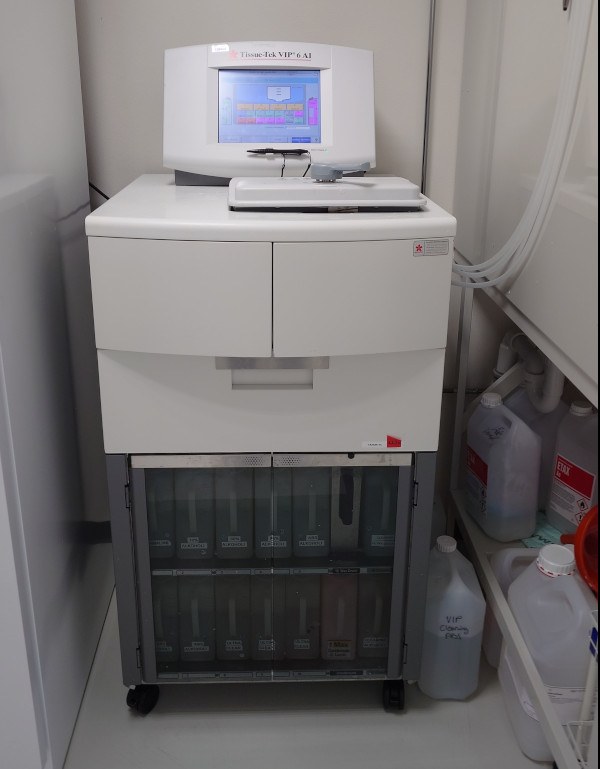
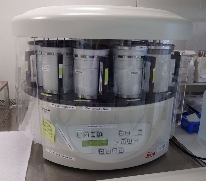
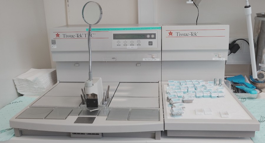
Tissue preparation
Histocore has two tissue processors for preparing paraffin blocks of solid tissue samples. Both formalin-fixed and Paxgene-fixed tissues can be prepared.
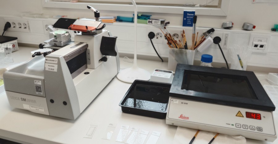
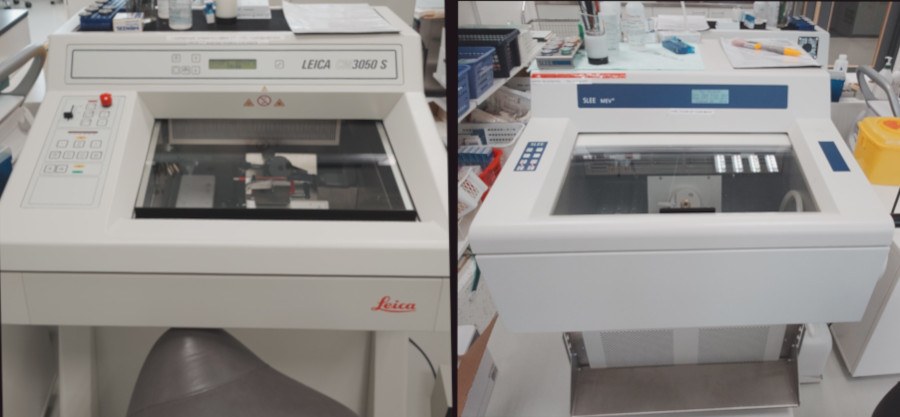
Histocore has two sliding and one rotary microtomes for cutting paraffin blocks (section thickness adjustable to 2-6 um). For cutting frozen sections Leica and Slee Mev+ cryomicrotomes are available.
Location: Arvo building, laboratory E325
Preparation of cultured cells
Users need to make preparations of cultured cells themselves. Cultured cells in suspension can be prepared as sectionable tissue blocks using the thrombin clotting technique. Alternatively, Sakura Cytocentrifuge can be used to prepare cell slides for IHC and IF staining.
Location: Arvo building, laboratory E325
Tissue staining
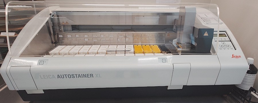
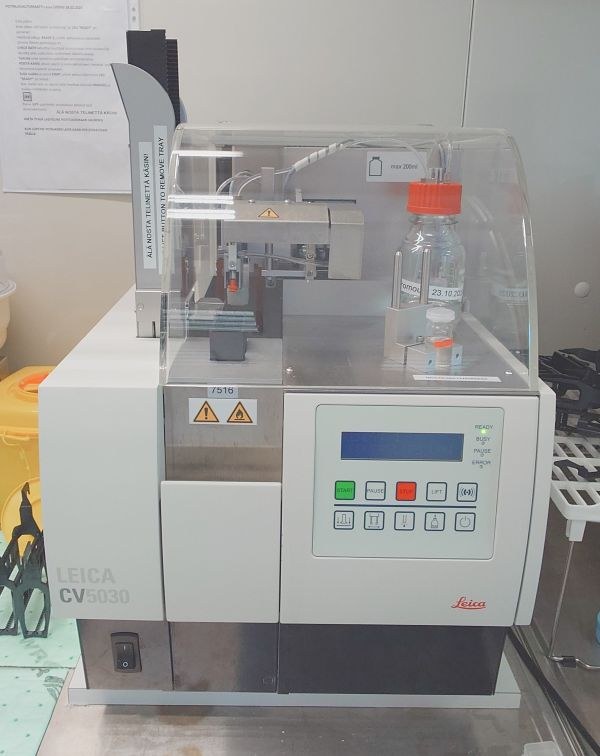
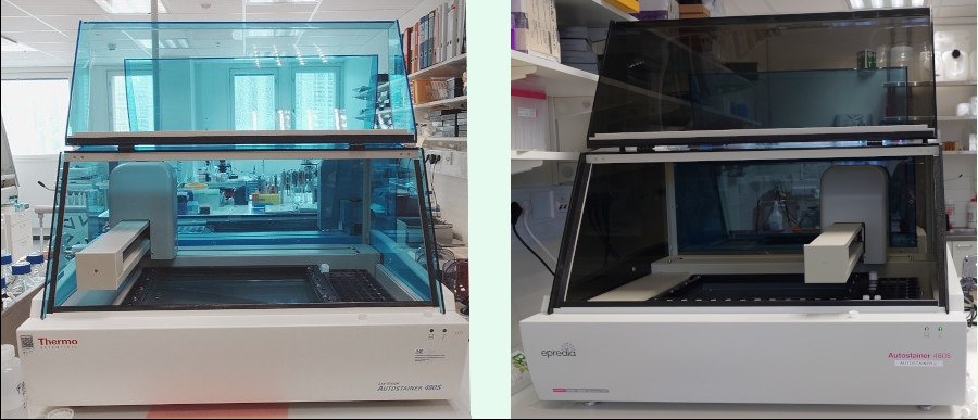
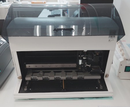
Histocore has Leica autostainer for automated hematoxylin-eosin and other routine stains. Slide coverslipping can be done automatically using DAKO coverslipper.
Location: Arvo building, laboratory E324
Immunohistochemistry
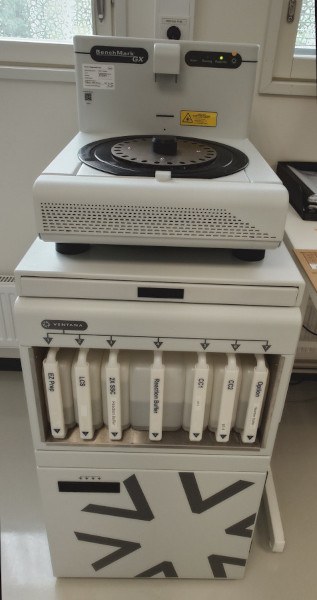
Histocore has one Ventana BenchMark and two LabVision autostainers for automated immunohistochemistry. Antigen retrieval can be done with a PT Module, Retriever 2100 or Biocare retriever, or a microwave oven using ph6 or pH9 buffers.
Location: Arvo building, laboratory E324
Immunofluorescence
Immunofluorescence staining can be done using LabVision Autostainer (see Immunohistochemistry section above) or manually using various staining racks available. Of these, JL-Stainer is well-suited for staining of 1-10 IF slides.
Location: Arvo building, laboratory E324
Tissue microarrays
Tissue microarray paraffin blocks can be prepared using the 3D Histech TMA Master. The system allows preparation of 0.6-2 mm diameter tissue cores punched and inserted in 1-2 recipient TMA blocks. Sectioning and staining of TMA blocks follows the principles of conventional tissue samples.
Location: Arvo building, laboratory E325
Light microscopes
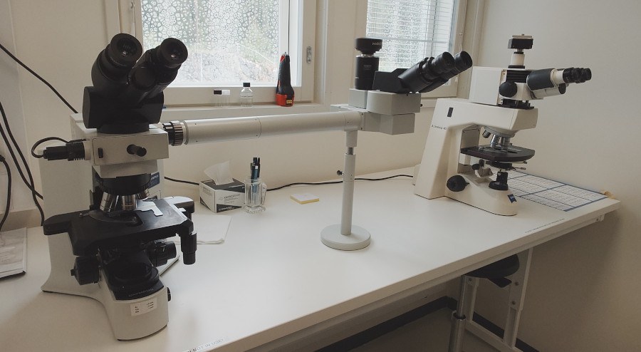
Histocore has several Olympus BX and Leica DM microscopes for slide inspection. Some of these are equipped with digital cameras for snapshot imaging.
Location: Arvo building, laboratory E324 and E325
Fluorescence microscope
Histocore has a Zeiss Axioplan microscope for multicolor fluorescence imaging and image capture. The microscope is equipped with ApoTome device, which allows structured illumination principle for generating sharp sections-within-section confocal-type images with all wavelengths. The microscope is operated with AxioVision software.
Laser capture microdissection
Veritas Arcturus Laser Capture system is available for isolation of cell islets or single cells from cell cultures and frozen tissue sections. The dissected material can be studied with molecular biology methods (e.g. PCR-based).
Location: Arvo building, laboratory E332
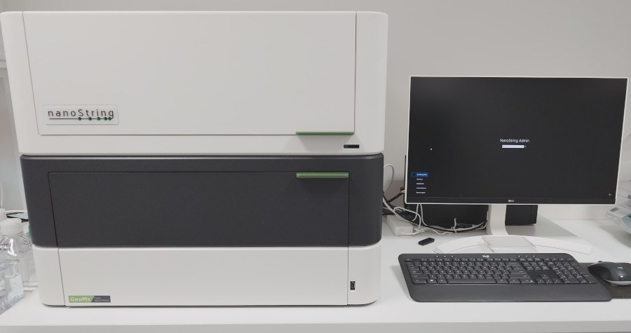
Digital Spatial Profiler
see GeoMx
Location: Arvo building, laboratory E326
Virtual microscopy
Virtual microscopy stands for acquiring and handling digitized versions of microscope glass slides, which are in turn commonly referred to as “virtual slides” or “whole-slide images (WSIs)”. Virtual slides are montage images consisting of up to thousands of high resolution digital micrographs.
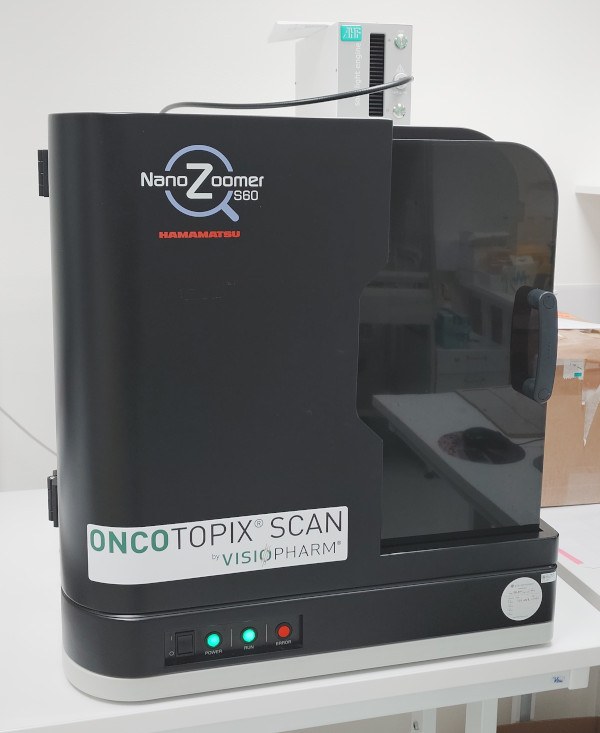
Virtual slide scanning systems
Histocore has two virtual slide scanners available, (Hamamatsu and Olympus), which enable scanning of microscope slides in batch. Alternatively, scanning of 2×3 inch macroslides is also possible. Scanning resolutions 0.31 um/pixel (using 10X lens) and 0.16 um/pixel (using 20X lens) can be selected by the user. Scanning with oil immersion lenses is also possible.
Both scanners allow scanning with epifluorescence illumination using blue, green and red emission wavelengths. The scanning output image format is jpeg2000.
Location: Arvo building, laboratory E322 (Hamamatsu), E332 (Olympus)
Virtual microscopy scanning system for microwell plates
Microwell plates can be scanned using a scanning system mounted in an inverted microscope (Olympus IX71) or using EVOS FL Auto. These systems allow scanning of cells grown on Petri dishes and 24-96 well microplates. The basic illumination system is phase contrast but fluorescent illumination (with motorized filter changer) is also possible.
Location: Arvo building, laboratory E332, E422, E462
The Tampere University Histology core facility ("Histocore") is available to the MET research community and by appointment to partners and contract research. Ask for further information if you have any questions!
After training by our staff, users can perform some of the basic histology laboratory tasks themselves. These include the use of microtomes, cryomicrotomes, tissue processors and microscopes, and the embedding of paraffin blocks.
Specialised equipment is only operated by histocore staff.
Technicians will assist you with technical questions and can perform tissue processing, staining and analysis for you whenever possible. Ask us about the services we have, also a full service is available!
Please note:
During the holiday season in July, the availability of the Histocore service will be limited. Self-service will remain available, but staff-assisted service will not be available until July 20. After that, staff-assisted service will be available on a limited basis until August 10.
Service work order
Please use the form below to request a service from Histocore:
Please complete the form carefully, ensuring that all relevant information is entered correctly. Unclear or incomplete information may delay the processing of samples. If necessary, ask Histocore staff for assistance. Email the completed form to histocore [at] tuni.fi (histocore(at)tuni.fi).
Note! Service Work Order is not required for self-service, but instrument booking is expected (see Booking System below)
Histocore has limited storage space. Please collect the samples yourself when the work is finished if you wish to keep them.
Booking system
All instruments run by users themselves must be booked using Agendo.
Please note that Histocore staff have priority access to the equipment.
Prices
(requires login to intranet)
For special projects ask for a quote!
Acknowledgement
All the users of the Tampere University histology core services are obligated to acknowledge the facility in their publications e.g. using the following phrase:
“The authors acknowledge the Tampere University Histocore Facility for their service.”
In addition, please add Tampere Histology facility as Facilities/Equipment for the publication in TUNICRIS.
Contact email for Tampere University Histocore services: histocore [at] tuni.fi (histocore(at)tuni.fi)