
Protein Services

Introduction
The facility offers services in protein expression, purification and characterization. The currently provided protein expression hosts (bacterial, insect and mammalian expression) cover a broad range of expression and post-translational modification requirements for a variety of life-science and industry needs. The produced recombinant proteins may be used in e.g. structural biology, protein-ligand and protein-protein interaction studies, and in the development of diagnostics.
Protein Expression Services
Expression vectors
The used expression vectors can either be provided by the customer or designed, case-by-case, by the Protein Service. The used vector backbone, included domains, fusion tags, linkers and protease cut-sites are selected based on the project needs.
E. coli protein expression
The Service carries a selection of E. coli strains for the expression of different types of proteins. For unknown proteins, we typically perform a small-scale expression test to confirm successful protein expression and to optimize expression parameters. After positive results, production-scale expression is performed using shake-flask cultures or small-scale fermentation system.
Protein Service offers protein production in bacterial hosts in up to 4.5 l batches using Labfors Infors 3 fermentor system. The accurately controlled growth conditions of the system enable higher cell densities and better product quality than what is achieved using conventional shake-flask cultures.
Insect cell expression using baculovirus system
The provided baculovirus expression service is based on the Bac-to-Bac system. Customer-provided or service-made donor plasmids are used to prepare 3-6 bacmids and the correct transposition of the gene-of-interest confirmed by PCR. Confirmed clones are transfected into the Sf9 or HighFive insect cells. Successful expression of the protein is typically confirmed at the level of secondary virus (P2, 50 ml culture volume) using Western blotting. Validated virus stocks are stable for up to one month and can be used both for further optimization of expression conditions and expression scale-up to 500 ml - 4.5 liter media volume.
Mammalian protein expression
The provided mammalian expression service is based on CHO and HEK 293 cell lines and covers both transient and stable protein expression. For most small-scale protein expression projects, transient expression provides the best balance between protein yield and resource use. For previously unknown proteins, a 25 ml pilot culture is initially used to confirm protein expression and solubility. Expression scale-up is possible up to 5-liter scale using parallel shake-flask cultures or Cytiva Wave bioreactor system.
Protein Isolation, Purification, and Characterization
Host cells and expression media are separated by centrifugation or filtration. For the purification of intracellular and periplasmic proteins, the cells are lysed using Emulsiflex high-pressure homogenizator, a probe sonicator or enzymatic methods. Protein isolation, purification and final polishing are typically done using different chromatographic methods (IMAC, IEX, SEC) ran using automated FPLC systems. When purification tag removal is necessary, specific proteases (e.g. TEV and 3C) can be used. Based on the project needs, quality of the final protein preparation can be evaluated using several methods, including analytical gel filtration, SDS-PAGE, 2D electrophoresis, dynamic light scattering, mass photometry, calorimetric methods and biosensor systems. The final product is provided to the customer either as a lyophilized powder or flash-frozen liquid in selected buffer system.
The Resources tab provides a brief summary of the tools and biophysical characterization instrumentation available.
Equipment
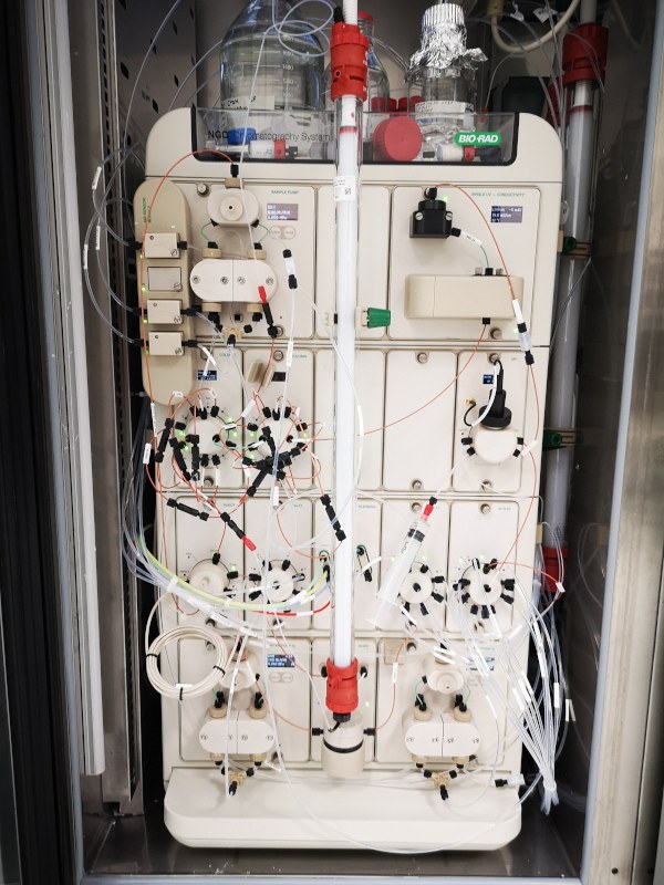
Chromatography systems. The facility has three complete ÄKTA chromatography systems installed for preparative chromatography work and they all include UV/VIS and conductivity detectors and are equipped with fraction collectors. One of the instruments is equipped with sample pump and pH electrode. We have also Shimadzu/Malver instrument especially suitable for analytical chromatography. This instrument has gradient pump, autosampler and fraction collector, and it is equipped with UV/VIS, fluorescence, DLS and SLS detectors.
Location:
Kauppi campus, Arvo building, laboratory E343
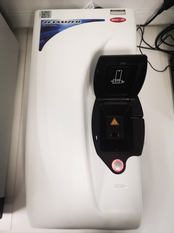
Dynamic light scattering (DLS) allows study of particle size from hydrodynamic diameter of 1 nm (size of sucrose molecule) and particle size distribution (homogeneity) in soluble samples. In addition, it is possible to measure Zeta potential which is a measure of the magnitude of the repulsion or attraction between particles. The significance of zeta potential is that it can be related to the stability of particle dispersions and allows one to find suitable conditions for samples (proteins, virus particles, nanoparticles etc). DLS is thus efficient method both in protein characterization and in protein formulation.
Location:
Kauppi campus, Arvo building, laboratory E301
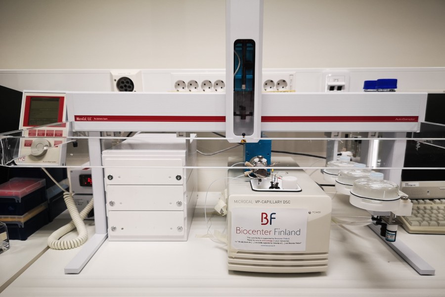
Differential scanning calorimetry (DSC) is primarily used to characterize stability and folding of macromolecules such as proteins. In addition, it can be used to screen protein-ligand interactions, because when a ligand preferentially binds to the native form of a protein, the protein is stabilized and the thermal transition midpoint (Tm) of the protein-ligand complex is higher than that of the protein in the absence of ligand. Therefore, DSC can be used for ligands with ultra-tight binding constants (1020 M-1) that cannot be measured by other methods, as well as a high throughput screening assay for drug discovery (up to 50 samples per day). The DSC instrument is equipped with refrigerated autosampler, enabling large sample sets.
Location:
Kauppi campus, Arvo building, laboratory E343
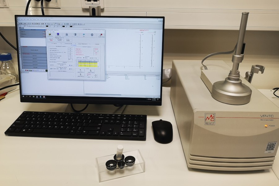
Isothermal titration calorimetry (ITC) is a thermodynamic technique that directly measures the heat released or absorbed during a biomolecular binding event. Measurement of this heat allows accurate determination of binding constants (KB), reaction stoichiometry (n), enthalpy (ΔH) and entropy (ΔS), thereby providing a complete thermodynamic profile of the molecular interaction in a single experiment. With ITC, it is possible to directly measure sub-millimolar to nanomolar binding constants (10-2 to 10-9 M) and nanomolar to picomolar binding constants (10-9 to 10-12 M) by using the competitive binding technique. Moreover, ITC is true in-solution technique and therefore no labeling or immobilization is required.
Location:
Kauppi campus, Arvo building, laboratory E343
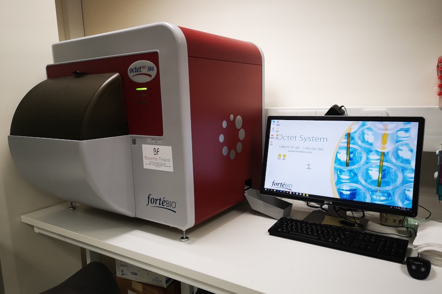
Biolayer interferometry (BLI) is technique, which is used to measure real-time, label-free analysis of biomolecular interactions and to provide information on affinity, kinetics and concentration. BLI technology monitors the binding of proteins and other biomolecules to their partners in real time. The binding interaction is continuously monitored by measuring the change in thickness of the protein layer on the biosensor tip. The method has no need to label the protein with fluorescent or chromogenic tags, thus eliminating interference issues. In addition, instrument uses 96 or 384 plates and 16 parallel sensors enabling high throughput assays.
Location:
Kauppi campus, Arvo building, laboratory E343
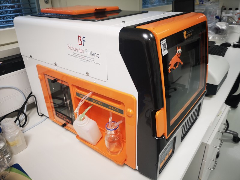
Surface plasmon resonance (SPR) use an optical method to measure the refractive index near a sensor surface in order to detect an interaction of one molecule that is immobilised onto the sensor’s surface. Method is used e.g. to search for binding partners or most commonly to measure kinetics of an interaction, i.e. the rates of complex formation (ka) and dissociation (kd). The previous parameters can be determined from the information in a sensorgram.
Location:
Kauppi campus, Arvo building, laboratory E301
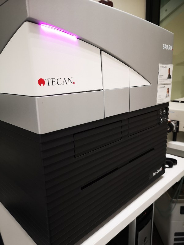
Multimode plate reader. Tecan Spark multimode plate reader can measure multiple different techniques including absorbance, luminescence, fluorescence intensity, fluorescence polarization, time-resolved fluorescence, FRET, as well as AlphaScreen, AlphaLISA and AlphaPlex. It can read microplates up to 1536 wells and has a temperature control from 18 to 42 °C. Instrument has both monochromators and filters allowing flexible wavelength selection with high measurement sensitivity.
Location:
Kauppi campus, Arvo building, laboratory E343
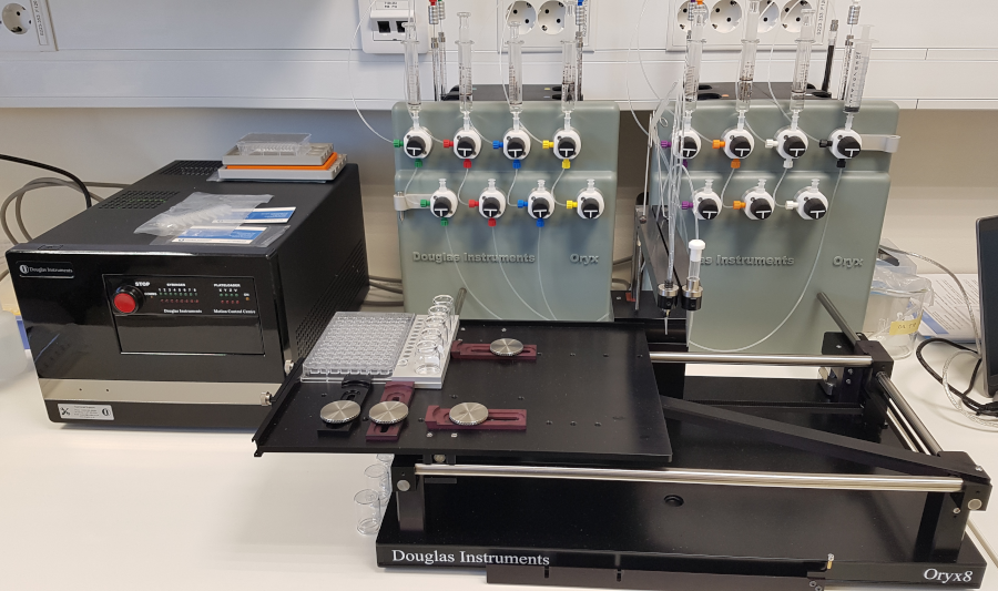
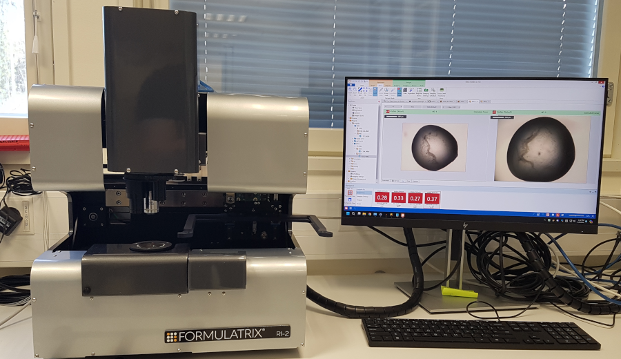
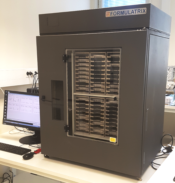
Protein crystallization. Protein crystallization facility is equipped with Oryx8 crystallization robot for setting up crystallization experiments and Formulatrix Rock Imagers RI2 and RI54 for imaging of crystallization plates. Crystallization screening can be done with sitting-drop and hanging-drop vapor-diffusion methods as well as microbatch under oil method in 96-well format. Commercial and in-house protein crystallization screens are available. Dedicated space at room temperature, 20 and 16 and 4 °C incubators are available for incubation of crystallization plates. Tools for crystal handling, freezing, and shipment to synchrotrons are available.
Location:
Kauppi campus, Arvo building, laboratory E327
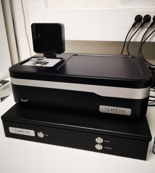
A mass photometer (MP) allows the precise determination of the mass of a single molecule in solution without the need for labels. The method is based on light scattering microscopy and can quickly and easily measure the mass of proteins, DNA/RNA molecules, macromolecules and even viruses. The measurement range of the Refey TwoMP instrument is 30 kDa - 5 MDa and the optimal sample concentration is 5 - 20 nM. Key applications besides mass determination are protein-protein interactions, DNA-protein interactions, protein oligomerisation, complex formation.
Location:
Kauppi campus, Arvo building, laboratory E343
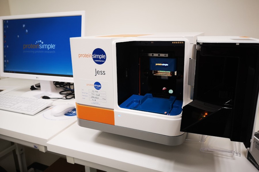
Protein Simple Jess is a compact protein analyzer that provides a fast and efficient way to analyze the quantity and qualitative characteristics of proteins. Jess uses capillary electrophoresis to separate and quantify proteins, enabling precise and reliable results with small sample amounts. Its automated functions make it an ideal tool for protein analysis in laboratories requiring fast and dependable results.
Location:
Kauppi campus, Arvo building, laboratory E223
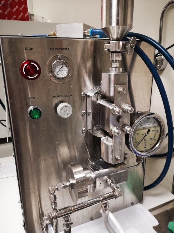
The Avestin Emulsiflex C3 is a lab scale, electric motor driven, high pressure homogeniser that rapidly reduces particles and droplets from micron to nanometer sizes.
Location:
Kauppi campus, Arvo building, laboratory E342
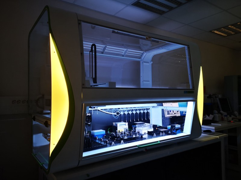
Revvity the JANUS® G3 automated liquid handling workstation offer flexible automated sample preparation solutions.
Location:
Kauppi campus, Arvo building, laboratory E216
Services provided
Protein Service facility offers services, expertise and hands-on guidance in recombinant protein production and analysis in life science research and drug discovery. The services offered by the facility cover the complete workflow from host selection and vector design to protein expression, purification and validation. Furthermore, we have a selection of biophysical protein characterization methods available to address diverse research questions in protein biology. The service is available for both academic and non-academic users.
The three currently available expression systems (bacteria, insect and mammalian cells) cover a large variety of different types of proteins and project requirements. Please contact the Protein Service team for assistance in protein expression host selection and project design. A typical workflow includes pilot scale protein production and purification, process scale-up, protein purification and protein characterization by various biochemical and biophysical methods, including light scattering methods and interaction assays.
Note: The Protein Service is equipped to handle BSL-1 materials only.
Getting started
To review your project requirements and get a service quotation, please contact the Protein Services team:
e-mail
proteinservices [at] tuni.fi (proteinservices[at]tuni[dot]fi)
Service coordinator:
Rolle Rahikainen, Ph.D.
rolle.rahikainen(at)tuni.fi
Tel: +358 50 3408919
Head of the facility:
Vesa Hytönen, Ph.D., Professor
vesa.hytonen(at)tuni.fi
Tel: +358 40 190 1517
Acknowledgement
All users are obliged to acknowledge the service provided by Tampere facility of Protein Services and the Biocenter Finland in their publications:
“The authors acknowledge the services provided by Tampere Protein Services and the Biocenter Finland”

Prices for academic users
| SERVICE | PRICE |
| E. coli expression and characterization | |
| Transformation per plasmid | 20 € |
| Production of a pre-culture from a tested clone | 20 € |
| 5 L Bacterial expression culture from pre-culture* | 300 € |
| Affinity Chromatography** | 150 € |
| Concentration and buffer change of protein sample | 25 € |
| SDS-PAGE with Coomassie staining | 50 € |
| SDS-PAGE with Oriole staining | 70 € |
| SDS-PAGE with Silver staining | 80 € |
| Immunoblotting*** | 100 € |
| ITC analysis per sample | 50 € |
| ITC equipment usage per day**** | 100 € |
| DSC analysis per sample (Minimum charge 3 samples) | 20 € |
| DLS analysis per sample | 20 € |
| SPR analysis per run*** | 50 € |
| Freeze drying per sample | 10 € |
| Octet BLI measurement (sensors do not include) | 25 € |
| BEVS | |
| Bacmid production (20 € x 6) | 120 € |
| Transfection Sf9 (HighFive 60 €) | 50 € |
| Optimization of the production by post infection follow up | 60 € |
| Pilot purification assay | 40 € |
| Scale up 1st 500 ml | 125 € |
| Scale up 2nd and following 500 mls | 75 € |
| Mammalian cell expression system | |
| Transfection and selection | 150 € |
| Cell adaptation | 100 € |
| Scale up 1 L | 200 € |
| Concentration | 350 € |
| Multimode plate reader | free |
| Protein crystallization | enquire |
The indicated pricing is for academic users, while non-academic users are invited to contact for a custom quotation.
Minimum invoicing 250 € / project.
Protein Service reserves the right to adjust pricing.
| * | Includes basic reagents and cell collection |
| ** | Affinity matrix will be charged separately |
| *** | Customer provides / pays for the antibody or SPR sensors |
| **** | User must attain tutoring for usage before starting experiments |
Prices for non-academic users
Please inquire!
General information
All stock products are recombinant proteins expressed in E. coli, except for Fibronectin, which is purified from human plasma. All proteins have been purified using one-step affinity chromatography and analyzed with SDS-PAGE. Protein concentration has been determined using UV/Vis spectroscopy. The proteins have been lyophilized and kept at -80 °C until shipment at ambient temperature.
Pricing and availability
The avidin proteins are sold at the following pricing:
- 500 µg 150€
- 1 mg 250 €
- 10 mg 1500 €
Please inquire for the availability and pricing of our Fibronectin products.
Protein products
AVIDIN PROTEINS
Tampere Protein Service carries a complete panel of recombinant avidin proteins suitable in diverse application is biosciences. Avidins are homotetrameric proteins with four identical subunits, each with one biotin binding site with extreme biotin binding affinity Kd ~ 10(-15M). Avidin contain one intra-chain disulfide bridges and no free sulfhydryl groups. Avidin expressed in E. coli is not glycosylated.
Recombinant avidin
SEQUENCE
- Recombinant avidin expressed in E. coli fermentation culture.
- Purification using 2-iminobiotin-Sepharose 4 Fast Flow resin.
- Avidin binds biotin with high affinity Kd ~ 10(-15M)
- Avidin binds 4-hydroxyazobenzene-2-carboxylic acid (HABA) with moderate affinity, resulting in a color change.
- The pI and extinction coefficient of recombinant Avidin are 9.69 and 23615 M-1 cm-1 (at 280 nm measured in water), respectively.
- Amino acid sequence: QTVARKCSLTGKW TNDLGSNMTI GAVNSRGEFT GTYITAVTAT SNEIKESPLH GTQNTINKRTQPTFGFTVNW KFSESTTVFT GQCFIDRNGK EVLKTMWLLR SSVNDIGDDW KATRVGINIFTRLRTQKE
REFERENCES
- Biotin induces tetramerization of a recombinant monomeric avidin. A model for protein-protein interactions. Laitinen OH, Marttila AT, Airenne KJ, Kulik T, Livnah O, Bayer EA, Wilchek M, Kulomaa MS. J Biol Chem. 2001 Mar 16;276(11):8219-24.
- Efficient production of active chicken avidin using a bacterial signal peptide in Escherichia coli. Hytönen VP, Laitinen OH, Airenne TT, Kidron H, Meltola NJ, Porkka EJ, Hörhä J, Paldanius T, Määttä JA, Nordlund HR, Johnson MS, Salminen TA, Airenne KJ, Ylä-Herttuala S, Kulomaa MS. Biochem J. 2004 384(Pt 2):385-90.
SWITCHavidin
- Switchable avidin variant.
- Switchavidin may be released permanently from biotinylated materials by applying a mixture of 2.5% citric acid and 0.25% SDS (2).
- The pI and extinction coefficient of Switchavidin are 7.02 and 23615 M-1 cm-1 (at 280 nm measured in water), respectively.
- Amino acid sequence: QTVARKCSLT GEWTNDLGSN MTIGAVNSRG EFTGTYITAV TATSNEIKES PLHGTQNTIN KRTQPTFGFT VNWKFSESTT VFTGQCFIDR NGKEVLKTHW LLRSSVNDIG DDWKATLVGI NIFTRLHTQE E
REFERENCES
- Biotin induces tetramerization of a recombinant monomeric avidin. A model for protein-protein interactions. Laitinen OH, Marttila AT, Airenne KJ, Kulik T, Livnah O, Bayer EA, Wilchek M, Kulomaa MS. J Biol Chem. 2001 Mar 16;276(11):8219-24.
- Reversible biofunctionalization of surfaces with a switchable mutant of avidin. Pollheimer P, Taskinen B, Scherfler A, Gusenkov S, Creus M, Wiesauer P, Zauner D, Schöfberger W, Schwarzinger C, Ebner A, Tampé R, Stutz H, Hytönen VP, Gruber HJ. Bioconjug Chem. 2013 Oct 16;24(10):1656-68
- Switchavidin: reversible biotin-avidin-biotin bridges with high affinity and specificity. Taskinen B, Zauner D, Lehtonen SI, Koskinen M, Thomson C, Kähkönen N, Kukkurainen S, Määttä JA, Ihalainen TO, Kulomaa MS, Gruber HJ, Hytönen VP. Bioconjug Chem. 2014 25(12):2233-43.
Chimeric avidin
- Chimera of chicken avidin and streptavidin with improved binding properties
- Contains one intra-chain disulfide bridge and no free sulfhydryl groups. Not glycosylated.
- The pI and extinction coefficient of chimeric avidin are 9.69 and 25105 M-1 cm-1 (at 280 nm measured in water), respectively.
- Amino acid sequence: QTVARKCSLT GKWTNDLGSN MTIGAVNSRG EFTGTYITAV ADNPGNITLS PLLGIQHKRA SQPTFGFTVN WKFSESTTVF TGQCFIDRNG KEVLKTMWLL RSSVNDIGDD WKATRVGYNI FTRLRTQKE
REFERENCES
- Biotin induces tetramerization of a recombinant monomeric avidin. A model for protein-protein interactions. Laitinen OH, Marttila AT, Airenne KJ, Kulik T, Livnah O, Bayer EA, Wilchek M, Kulomaa MS. J Biol Chem. 2001 Mar 16;276(11):8219-24.
- Chimeric Avidin--NMR structure and dynamics of a 56 kDa homotetrameric thermostable protein. Tossavainen H, Kukkurainen S, Määttä JA, Kähkönen N, Pihlajamaa T, Hytönen VP, Kulomaa MS, Permi P. PLoS One. 2014 Jun 24;9(6):e100564.
- Structural and functional characteristics of chimeric avidins physically adsorbed onto functionalized polythiophene thin films. Albers WM, Pelto JM, Suspène C, Määttä JA, Yassar A, Hytönen VP, Vikholm-Lundin IM, Tappura K. ACS Appl Mater Interfaces. 2012 Aug;4(8):4067-77.
- Chimeric avidin shows stability against harsh chemical conditions--biochemical analysis and 3D structure. Määttä JA, Eisenberg-Domovich Y, Nordlund HR, Hayouka R, Kulomaa MS, Livnah O, Hytönen VP. Biotechnol Bioeng. 2011 Mar;108(3):481-90.
- Neutralized chimeric avidin binding at a reference biosensor surface. Ray S, Steven RT, Green FM, Höök F, Taskinen B, Hytönen VP, Shard AG. Langmuir. 2015 31(6):1921-30.
Charge-neutralized chimeric avidin
- Chimeric avidin with charge-neutralizing mutations
- The pI and extinction coefficient of CHARGE-NEUTRALIZED CHIMERIC AVIDIN are 6.92 and 25105 M-1 cm-1 (at 280 nm measured in water), respectively.
- Amino acid sequence: QTVARKCSLT GEWTNDLGSN MTIGAVNSNG EFTGTYITAV ADNPGNITLS PLLGIQHKRA SQPTFGFTVN WKFSESTTVF TGQCFIDRNG KEVLKTMWLL RSSVNDIGDD WKATRVGYNI FTRLHTQEE
REFERENCES
- Biotin induces tetramerization of a recombinant monomeric avidin. A model for protein-protein interactions. Laitinen OH, Marttila AT, Airenne KJ, Kulik T, Livnah O, Bayer EA, Wilchek M, Kulomaa MS. J Biol Chem. 2001 Mar 16;276(11):8219-24.
- Chimeric Avidin--NMR structure and dynamics of a 56 kDa homotetrameric thermostable protein. Tossavainen H, Kukkurainen S, Määttä JA, Kähkönen N, Pihlajamaa T, Hytönen VP, Kulomaa MS, Permi P. PLoS One. 2014 Jun 24;9(6):e100564.
- Structural and functional characteristics of chimeric avidins physically adsorbed onto functionalized polythiophene thin films. Albers WM, Pelto JM, Suspène C, Määttä JA, Yassar A, Hytönen VP, Vikholm-Lundin IM, Tappura K. ACS Appl Mater Interfaces. 2012 Aug;4(8):4067-77.
- Chimeric avidin shows stability against harsh chemical conditions--biochemical analysis and 3D structure. Määttä JA, Eisenberg-Domovich Y, Nordlund HR, Hayouka R, Kulomaa MS, Livnah O, Hytönen VP. Biotechnol Bioeng. 2011 Mar;108(3):481-90.
- Neutralized chimeric avidin binding at a reference biosensor surface. Ray S, Steven RT, Green FM, Höök F, Taskinen B, Hytönen VP, Shard AG. Langmuir. 2015 31(6):1921-30.
Charge-neutralized chimeric avidin with linkable C-terminal cysteine
- Charge-neutralized chimeric avidin with a free C-terminal sulfhydryl.
- Allows avidin conjugation with sulfhydryl reactive chemistries (e.g. Maleimide, iodoacetamide etc.)
- The pI and extinction coefficient of Charge-neutralized chimeric avidin with linkable C-terminal cysteine are 6.91 and 25105 M-1 cm-1 (at 280 nm measured in water), respectively.
- Amino acid sequence: QTVARKCSLT GEWTNDLGSN MTIGAVNSNG EFTGTYITAV ADNPGNITLS PLLGIQHKRA SQPTFGFTVN WKFSESTTVF TGQCFIDRNG KEVLKTMWLL RSSVNDIGDD WKATRVGYNI FTRLHTQEEC
REFERENCES
- Biotin induces tetramerization of a recombinant monomeric avidin. A model for protein-protein interactions. Laitinen OH, Marttila AT, Airenne KJ, Kulik T, Livnah O, Bayer EA, Wilchek M, Kulomaa MS. J Biol Chem. 2001 Mar 16;276(11):8219-24.
- Chimeric Avidin--NMR structure and dynamics of a 56 kDa homotetrameric thermostable protein. Tossavainen H, Kukkurainen S, Määttä JA, Kähkönen N, Pihlajamaa T, Hytönen VP, Kulomaa MS, Permi P. PLoS One. 2014 Jun 24;9(6):e100564.
- Structural and functional characteristics of chimeric avidins physically adsorbed onto functionalized polythiophene thin films. Albers WM, Pelto JM, Suspène C, Määttä JA, Yassar A, Hytönen VP, Vikholm-Lundin IM, Tappura K. ACS Appl Mater Interfaces. 2012 Aug;4(8):4067-77.
- Chimeric avidin shows stability against harsh chemical conditions--biochemical analysis and 3D structure. Määttä JA, Eisenberg-Domovich Y, Nordlund HR, Hayouka R, Kulomaa MS, Livnah O, Hytönen VP. Biotechnol Bioeng. 2011 Mar;108(3):481-90.
- Neutralized chimeric avidin binding at a reference biosensor surface. Ray S, Steven RT, Green FM, Höök F, Taskinen B, Hytönen VP, Shard AG. Langmuir. 2015 31(6):1921-30.
HUMAN FIBRONECTIN
STRUCTURE:
Fibronectin is a major constituent of connective tissue and plasma. Fibronectin exists as a protein dimer, consisting of two nearly identical monomers linked by a pair of disulfide bonds. The fibronectin protein is produced from a single gene, but alternative splicing of its pre-mRNA leads to the creation of several isoforms. Fibronectin binds cell surface receptors and various compounds including collagen, fibrin, heparin, DNA, and actin. Fibronectin is involved in cell adhesion, cell motility, opsonization, wound healing, and maintenance of cell shape. It is also involved in osteoblast compaction through the fibronectin fibrillogenesis cell-mediated matrix assembly process, and is essential for osteoblast mineralization.
Two types of fibronectin are present in vertebrates:
- soluble plasma fibronectin (formerly called "cold-insoluble globulin", or CIg) is a major protein component of blood plasma (300 μg/ml) and is produced in the liver by hepatocytes.
- insoluble cellular fibronectin is a major component of the extracellular matrix. It is secreted by various cells, primarily fibroblasts, as a soluble protein dimer and is then assembled into an insoluble matrix in a complex cell-mediated process.
Molecular weight of isoform 1 of fibronectin is 262,625 Da based on amino acid sequence. The observed size in reducing SDS-PAGE is around 250 kDa.
PHYSICAL PROPERTIES:
Calculated isoelectic point (pI) of FN is 5.5-6.3.
Extinction coefficient of FN is 367375 M-1 cm-1 (at 280 nm measured in water).
METHOD OF PREPARATION:
Fibronectin is a major protein found in human plasma and purified by affinity chromatography using Gelatin Sepharose 4B (GE Healthcare Life Sciences). The purified protein is lyophilized in 150 mM NaCl, 10 mM sodium phosphate pH 7.5 and 1% trehalose to ensure stability in storage. Purity is over 95% based on SDS-PAGE (band around 250 kDa).
SOLUBILITY / SOLUTION STABILITY:
Fibronectin is shipped at ambient temperature. Upon arrival, store dry at –20°C or lower. Lyophilized product is moisture-sensitive. Reconstituted and sterile-filtered product can be stored for 1 month at +2...8°C.
PRODUCT DESCRIPTION / USAGE:
Reconstitution of lyophilized fibronectin is done by using 1 ml of warm (37°C) PBS per 1.4 mg of lyophilized fibronectin. Incubate at 37°C for 3 hours, occasionally inverting the vial. Do not vortex. Reconstituted solution contains 1 mg/ml of soluble fibronectin and a minor quantity of insoluble fibronectin aggregate. Filter slowly through prewetted 0.2 µm membrane filter. Concentration 1 mg/ml of fibronectin corresponds to A280 - 1.4
REFERENCES
- Fibronectin at a glance. Pankov R, Yamada KM. Journal of Cell Science 115 (Pt 20): 3861–3. doi:10.1242/jcs.00059.
- Talin-bound NPLY motif recruits integrin-signaling adapters to regulate cell spreading and mechanosensing. Pinon P, Pärssinen J, Vazquez P, Bachmann M, Rahikainen R, Jacquier MC, Azizi L, Määttä JA, Bastmeyer M, Hytönen VP, Wehrle-Haller B. J Cell Biol. 2014,205(2):265-81.
Contacts
e-mail
proteinservices [at] tuni.fi (proteinservices[at]tuni[dot]fi)
Service coordinator:
Rolle Rahikainen, Ph.D.
rolle.rahikainen(at)tuni.fi
Tel: +358 50 3408919
Head of the facility:
Vesa Hytönen, Ph.D., Professor
vesa.hytonen(at)tuni.fi
Tel: +358 40 190 1517
Room: ARVO F331
Street address:
Arvo Ylpön katu 34
33520 Tampere
Finland
Reference Publications
Progression of herpesvirus infection remodels mitochondrial organization and metabolism
Simon Leclerc, Visa Ruokolainen, Alka Gupta, Axel Ekman, Jian-Hua Chen, Sergey Kapishnikov, Eric Dufour, Vesa Hytonen, Eva Pereiro, Tony McEnroe, Kenneth Fahy, Carolyn Larabell, Venera Weinhardt, Vesa Aho, Maija Vihinen-Ranta. Biophysical Journal 123 (3), 521a. 2024
SpyTag/SpyCatcher display of influenza M2e peptide on norovirus-like particle provides stronger immunization than direct genetic fusion
Vili Lampinen, Stina Gröhn, Saana Soppela, Vesna Blazevic, Vesa P Hytönen, Minna M Hankaniemi. Frontiers in Cellular and Infection Microbiology 13, 1216364. 2023
Structural mechanism for inhibition of PP2A-B56α and oncogenicity by CIP2A
Karolina Pavic, Nikhil Gupta, Judit Domènech Omella, Rita Derua, Anna Aakula, Riikka Huhtaniemi, Juha A Määttä, Nico Höfflin, Juha Okkeri, Zhizhi Wang, Otto Kauko, Roosa Varjus, Henrik Honkanen, Daniel Abankwa, Maja Köhn, Vesa P Hytönen, Wenqing Xu, Jakob Nilsson, Rebecca Page, Veerle Janssens, Alexander Leitner, Jukka Westermarck. Nature Communications 14 (1), 1143. 2023
Biochemical and Biophysical Characterization of Carbonic Anhydrase VI from Human Milk and Saliva
Alma Yrjänäinen, Maarit S Patrikainen, Latifeh Azizi, Martti EE Tolvanen, Mikko Laitaoja, Janne Jänis, Vesa P Hytönen, Alessio Nocentini, Claudiu T Supuran, Seppo Parkkila. The Protein Journal 41 (4-5), 489-503. 2022
Hepcidin is potential regulator for renin activity
Jaakko Piesanen, Jarkko Valjakka, Sanna Niemelä, Marjut Borgenström, Seppo Nikkari, Vesa Hytönen, Juha Määttä, Tarja Kunnas. Plos one 17 (4), e0267343. 2022
Stable immobilisation of His-tagged proteins on BLI biosensor surface using cobalt
Auer S, Azizi L, Faschinger F, Blazevic V, Vesikari T, Gruber HJ, Hytönen VP. Sensors and Actuators B: Chemical. 2017 May 243:104-113.
Structural characterization of core-bradavidin in complex with biotin
Agrawal N, Määttä JAE, Kulomaa MS, Hytönen VP, Johnson MS, Airenne TT. PLoS One. 2017 Apr 20;12(4):e0176086
Transglutaminase 2-specific coeliac disease autoantibodies induce morphological changes and signs of inflammation in the small-bowel mucosa of mice
Kalliokoski S, Piqueras VO, Frías R, Sulic AM, Määttä JA, Kähkönen N, Viiri K, Huhtala H, Pasternack A, Laurila K, Sblattero D, Korponay-Szabó IR, Mäki M, Caja S, Kaukinen K, Lindfors K. Amino Acids. 2017 Mar;49(3):529-540
Artificial Avidin-Based Receptors for a Panel of Small Molecules. Lehtonen SI, Tullila A, Agrawal N, Kukkurainen S, Kähkönen N, Koskinen M, Nevanen TK, Johnson MS, Airenne TT, Kulomaa MS, Riihimäki TA, Hytönen VP. ACS Chem Biol. 2016 11(1):211-21
The Minor Capsid Protein VP11 of Thermophilic Bacteriophage P23-77 Facilitates Virus Assembly by Using Lipid-Protein Interactions.
Pawlowski A, Moilanen AM, Rissanen IA, Määttä JA, Hytönen VP, Ihalainen JA, Bamford JK. J Virol. 2015 Aug;89(15):7593-603